How Common Is Bad News At 20 Week Scan- All You Need To Know
How common is bad news at 20 week scan? Being expectant parents, the 20-week scan is often one of the most highly anticipated moments in a pregnancy journey.
It’s a time when you finally get to see your beautiful baby’s tiny fingers and toes, hear their melodious heartbeat, and confirm that everything is advancing as it should be!
However, for some parents, this joyous occasion can become a nightmare when an abnormality is detected during the 20 week anomaly scan. The fear of receiving bad news at the 20 week ultrasound is a common concern among many anticipating parents, and understandably so.
But just how common is bad news at 20 week scan?
In this article, we’ll take a closer look at the chances of abnormality at 20 week scan, the types of abnormalities that may be detected, and what steps can be taken if an abnormal 20 week ultrasound is noticed.
We understand that this can be a stressful and emotional time, and our goal is to provide the necessary knowledge and support to help guide you through this process!
Further ahead, we’ll discuss:
- What is a 20 week anatomy scan?
- How common is bad news at 20 week scan?
- What to expect at 20 week ultrasound?
- What are some birth defects detected at 20 week ultrasound?
- What are the chances of abnormality at 20-week scan?
- 9 Questions to ask at 20 week scan!
Without further ado, let’s begin!
Related Article—All you need to know about pregnancy stretch marks!
What is a 20 week anatomy scan?
A 20 week anatomy scan is a detailed ultrasound examination that is typically done during the second trimester of pregnancy!
This scan is also known as a mid-pregnancy ultrasound or a level 2 ultrasound. It is performed to assess the evolution of the fetus and to check for any potential irregularities.
1. When is an anatomy scan done?
The anatomy scan for pregnancy is usually done between 18 to 22 weeks of pregnancy, as this is when the baby’s anatomy has developed enough to be visible on the ultrasound.
This is a critical time in the baby’s development, and the scan can provide crucial information about the baby’s growth and well-being!
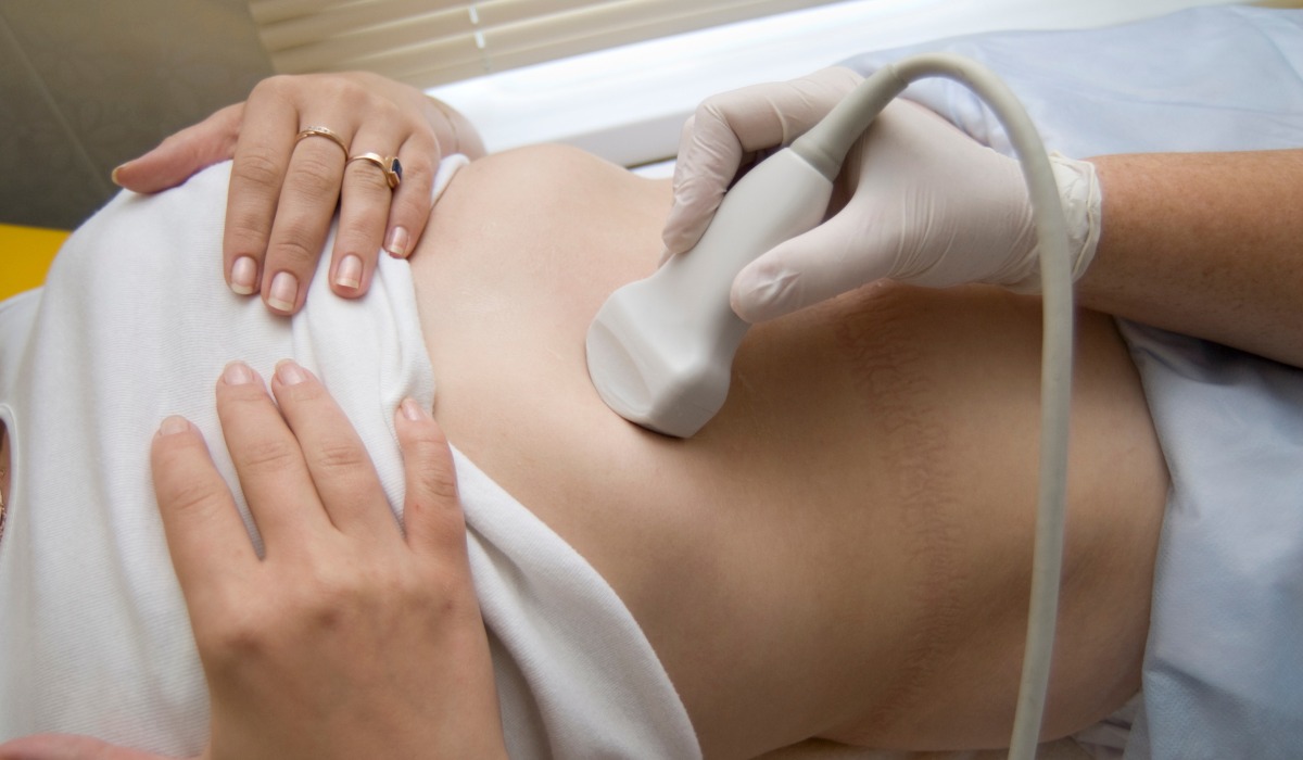
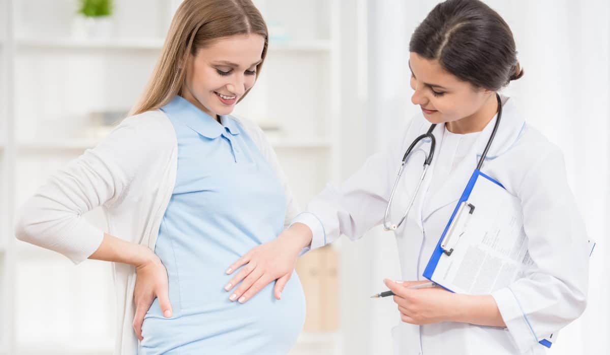
2. How to prepare for 20 week ultrasound?
Before the anomaly scan, your healthcare expert or doctor will likely provide you with instructions on how to prepare for 20 week ultrasound.
This may include drinking water or other fluids to ensure that your bladder is full, as this can help deliver better images. It’s also important to wear comfortable clothing and avoid any lotions or creams on your belly, as this can interfere with the 20 week ultrasound pictures!
Related Article—How to prepare for baby without missing anything out?
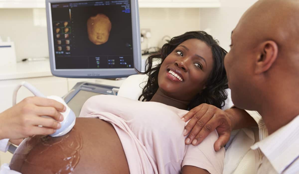
3. What happens at an anatomy scan?
During the 20 week ultrasound anatomy scan, a trained technician or sonographer will use a handheld device (called a transducer) to send sound waves into your uterus.
These sound waves bounce off the developing fetus and create images on a screen, which the technician can use to evaluate the baby’s anatomy.
The technician will typically start by measuring the baby’s head, abdomen, and femur bone to study growth and estimate the baby’s size. They will also check the baby’s organs, including the heart, brain, lungs, kidneys, and bladder, to ensure that they are developing properly.
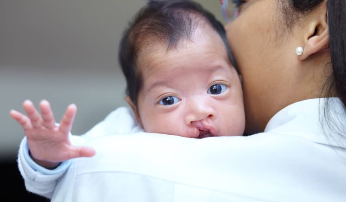
Moreover, the 20 week pregnancy scan can also detect other potential issues, such as cleft lip or palate, neural tube defects, and limb abnormalities!
4. How long does a 20 week ultrasound take?
The 20-week anatomy scan can take anywhere from 30 minutes to an hour, depending on the complexity of the exam and the position of the baby!
During the 20 week sonogram, the technician may ask you to change positions or move around to get better photographs. You may also be able to see the insider visions on a screen and ask questions about what you’re seeing.
5. What does a 20 week ultrasound look like?
At 20 weeks, the fetus is well-developed and the ultrasound can show many details about its anatomy. Some of the features that may be visible on the ultrasound include:
- The head, brain, and face, including the eyes, nose, and mouth
- The arms and legs, including the fingers and toes
- The spine and ribs
- The heart and major blood vessels
- The stomach, kidneys, and bladder
- The sex of the fetus (if the parents want to know)
The images from the ultrasound will appear in black and white and may be difficult to interpret without the help of a trained technician. Your healthcare expert will be able to explain the images to you and answer any questions you may have about your baby’s development.
6. What to expect at anatomy scan?
After the 20 week anatomy scan 3D, you will normally receive a report with the results. Your healthcare expert will review it with you later. If any abnormalities are detected, you may be referred to a specialist for further testing or monitoring.
Overall, the 20 week scan pregnancy is an important part of prenatal care, providing valuable information about the baby’s growth. It helps ensure a healthy pregnancy.
While it can be nerve-wracking to wait for the results, it’s noteworthy to remember that most babies are born healthy and that the vast majority of 20-week scans are normal.
Still, it’s only natural to wonder about the chances of abnormality at 20-week scan. You may wonder, “How common is bad news at 20 week scan?” Let’s try to figure it out together!
How common is bad news at 20 week scan?
The 20 week fetus ultrasound is a critical element of prenatal care, providing significant data about the fetus’s maturation. Furthermore, it also helps in identifying any potential issues that may require further evaluation or treatment!
While the majority of 20-week scans reveal that the fetus is healthy, unfortunately, there are instances where the scan detects abnormalities or potential health concerns for the baby!
Some of the most common abnormalities detected during a 20-week scan include structural problems with the heart or brain, as well as issues with the spine or kidneys.
What to expect at 20 week ultrasound? Here are a couple of anomaly scan results explained:
– According to a study published in PubMed Central, out of 4080 pregnant women undergoing ultrasound, 312 (7.6%) had fetal structural malformation.
– Another research found that approximately 2 to 3% of all pregnancies will have a major structural fetal anomaly detected on the 20-week scan.
This means that while it is not incredibly common for a serious issue to be detected, it is still a possibility that expectant parents should be aware of.
It’s essential to note that a 20-week scan is not a definitive diagnosis of any problems, but rather an indication that further testing may be required.
In some cases, a follow-up scan or additional tests may be recommended to get a clearer picture of the situation and determine the best course of action.
Related Article—10 baby registry checklist must haves!
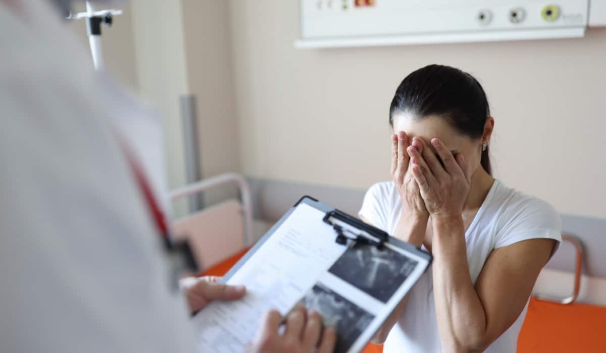
What happens at 20 week appointment?
Receiving bad news during a 20-week scan can be a difficult and emotional experience for expecting parents. However, you must remember that there are support systems available, such as genetic counselors and perinatal care teams, who can provide you with proper guidance throughout the process!
Overall, while bad news is not common during a 20-week scan, it is still a possibility. This is why, expecting parents need to be aware of the potential risks and prepared for any outcomes.
What abnormalities can be detected on an ultrasound?
If you’re wondering, “How common is bad news at 20 week scan?” you must also be wary of what the bad news brings!
The 20-week anatomy scan is a comprehensive ultrasound examination that is generally performed during the second trimester of pregnancy.
This scan is designed to evaluate the baby’s all-around health and development, by providing a detailed look at the baby’s bones, heart, brain, spinal cord, face, kidneys, and abdomen.
One of the primary objectives of the 20-week scan is to screen for certain rare conditions, which may not be detectable by other means.
In total, the 20 weeks ultrasound scan is capable of detecting 11 specific conditions, including neural tube defects, heart defects, abdominal wall defects, and others. By identifying these conditions early on, medical professionals can take suitable steps to manage or treat them and help ensure the best possible outcomes for both the mother and the baby.
Here’s a list of 20 week ultrasound abnormalities that may be discovered in a normal 20 week ultrasound:
1. Anencephaly
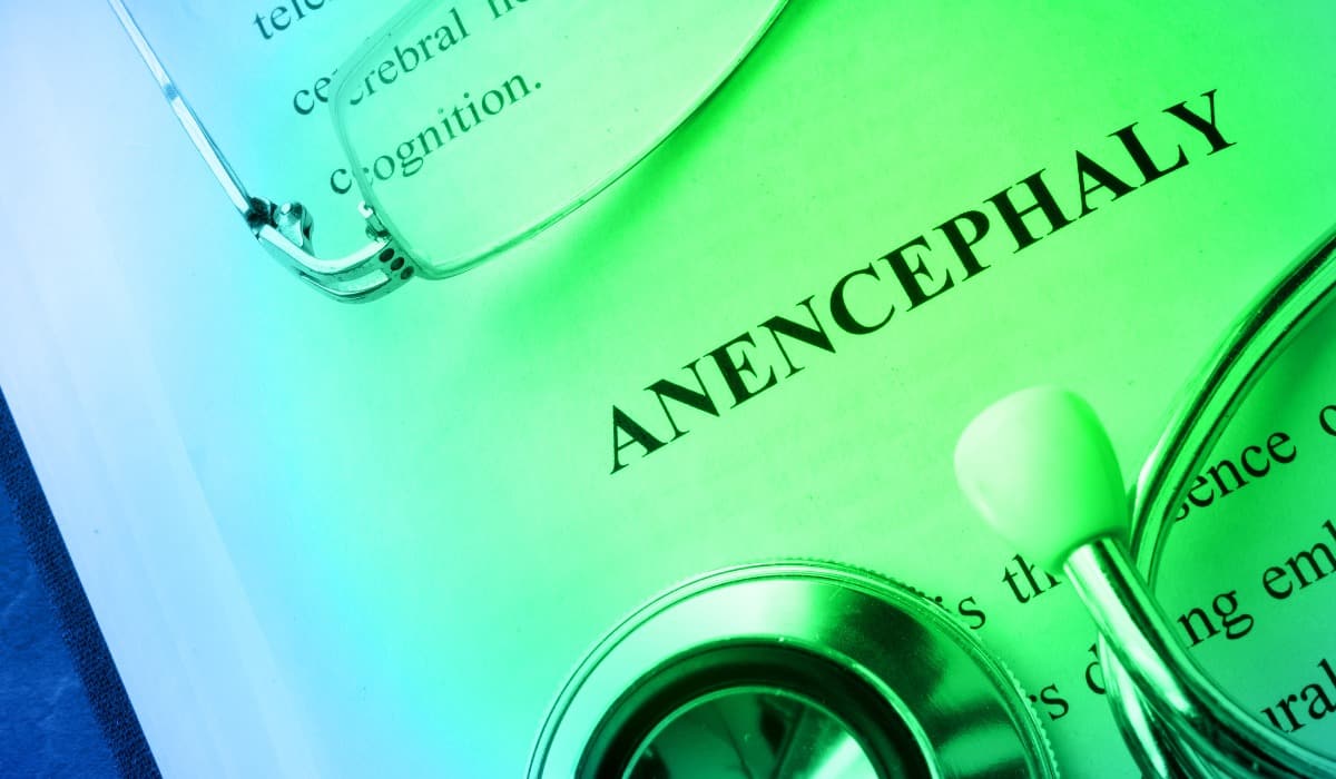
(Affects approximately 1 in 5,000 live births in the United States)
Anencephaly is a serious birth defect in which a baby is born without parts of the brain and skull. Babies with anencephaly usually do not survive for very long after birth, and there is currently no cure or treatment for this condition.
The exact cause of anencephaly is not known, but it is believed to be related to a combination of genetic and environmental factors.
Conceiving women or women who are pregnant can reduce their risk of having a baby with anencephaly by taking certain precautions. This includes getting enough folic acid and avoiding certain medications and environmental toxins
Related Article—Is Coffee Safe during Pregnancy?
2. Open Spina Bifida
(Affects 1 in 2,858 live births in the United States)
Open spina bifida, also known as myelomeningocele, is a birth defect that occurs when the spinal cord and the surrounding nerves and tissues protrude through an opening in the baby’s back.
This can lead to a range of complications, including paralysis, bowel and bladder problems, and hydrocephalus (an accumulation of fluid in the brain).
Open spina bifida is usually diagnosed during pregnancy or shortly after birth. The treatment for this health issue may involve surgery to repair the defect and prevent further damage to the spinal cord.
3. Diaphragmatic hernia
(Affects 1 in 2,500 to 5,000 live births in the United States)
Diaphragmatic hernia is a congenital birth defect that affects the formation of the diaphragm, a large, dome-shaped muscle that separates the chest cavity from the abdominal cavity.
The diaphragm is responsible for controlling breathing by expanding and contracting the lungs.
In babies with a diaphragmatic hernia, the diaphragm has not formed properly, leaving a hole that allows the organs from the abdomen to move up into the chest cavity. This can put pressure on the lungs, heart, and other organs, leading to a range of potential difficulties.
Diaphragmatic hernia is a relatively rare condition, affecting approximately 1 in 2,500 live births. It is more common in males than females and occurs more frequently on the left side of the body.
In some cases, the diaphragmatic hernia may be detected during a routine ultrasound at 20 weeks pregnant, allowing for early diagnosis and planning.
Infants with more severe cases of diaphragmatic hernia may experience difficulty breathing, rapid breathing, blue-tinged skin, a rapid heart rate, and other signs of respiratory distress. In some cases, the condition may be accompanied by other birth defects or genetic abnormalities.
4. Gastroschisis
(Affects 1 in 4,000 live births in the United States)
Gastroschisis is a congenital birth defect that impacts the abdominal wall of developing fetuses. This condition occurs when the abdominal wall fails to close completely, resulting in an opening or hole in the abdomen.
As a result, the intestines and other organs pierce through the hole and are exposed outside of the body!
Some of the risk factors for developing gastroschisis include maternal age under 20, smoking during pregnancy, and the use of certain medications or substances.
The birth defect is commonly diagnosed with a fetal ultrasound 20 weeks, which can show the exposed organs outside of the fetal abdomen. Treatment for gastroschisis typically involves the delivery of the baby by cesarean section and immediate surgical repair of the defect.
The exposed organs are gently placed back into the abdominal cavity and the wall is closed surgically.
Babies born with gastroschisis may experience complications such as intestinal damage, infection, and difficulty absorbing nutrients. They may require specialized care in a neonatal intensive care unit (NICU) until they are stable enough to go home.
With fast and appropriate medical care, many babies born with gastroschisis can recover fully and go on to live healthy, normal lives. However, the condition can be life-threatening in severe cases or when left untreated.
5. Cleft lip
(Affects 1 in 940 live births worldwide)
Cleft lip is a birth defect that affects the formation of the upper lip of your baby. It occurs when the tissues that form the lip do not join together properly during fetal growth.
This can result in a split or opening in the lip, which can range in severity from a small notch to a complete separation that extends into the nose.
A cleft lip can occur alone or in combination with a cleft palate, which affects the roof of the mouth. Treatment for cleft lip usually involves surgery to restore the defect, and may also involve speech therapy, dental care, and other supportive therapies!
6. Exomphalos
(Affects approximately 1 in 5,000 live births worldwide)
Another similar birth defect that affects the abdominal wall of a baby is Exomphalos. Also known as omphalocele, this is a condition that occurs when the muscles of the abdominal wall do not close properly.
The birth defect allows some of the abdominal organs, such as the intestines, liver, or spleen, to protrude through a hole in the belly button area and be covered by a thin sac.
Some of the risk factors for developing exomphalos include maternal age over 35, maternal obesity, and maternal smoking.
7. Serious Cardiac Abnormalities

(Affects 8 out of every 1,000 babies born worldwide)
Serious cardiac abnormalities refer to any structural defects or functional disorders of the heart that affect its ability to function properly!
These abnormalities can occur in babies during pregnancy and may lead to serious health complications, and even death, if not diagnosed and treated ASAP.
There are several types of serious cardiac abnormalities that can affect babies, including:
– Hypoplastic left heart syndrome: a rare condition where the left side of the heart is underdeveloped, making it difficult for the heart to pump blood to the body.
– Tetralogy of Fallot: a combination of four heart defects that affect the flow of blood through the heart, causing oxygen-poor blood to circulate through the body.
– Transposition of the great arteries: a condition where the two main arteries that carry blood out of the heart are reversed, causing oxygen-poor blood to be pumped to the body instead of the lungs.
– Truncus arteriosus: a condition where the main arteries that carry blood out of the heart fail to separate during fetal development. This leads to a mixture of oxygen-poor and oxygen-rich blood.
– Total anomalous pulmonary venous connection: a condition where the veins that carry oxygen-rich blood from the lungs to the heart are abnormally connected to the heart, causing oxygen-poor blood to circulate through the body.
Serious cardiac abnormalities are usually diagnosed with a fetal echocardiogram, a specialized ultrasound that provides detailed images of the fetal heart. Treatment for serious cardiac abnormalities may involve medication, surgery, or a combination of both.
Babies born with serious cardiac abnormalities may need ongoing medical follow-up and monitoring to ensure that their heart function remains stable.
What are the chances of no heartbeat at 20 week scan?
The chance of no heartbeat at 20 week scan is relatively low!
According to the American College of Obstetricians and Gynecologists (ACOG), the fetal heartbeat should be visible and audible on ultrasound by the 8th week of pregnancy. Plus, it should be easily detectable by 12 weeks. By the 20th week, the heartbeat is usually strong and relatively easy to detect.
However, in rare cases, a 20-week ultrasound may not be able to detect a fetal heartbeat. This could be due to several reasons, such as a miscarriage or a fetal abnormality that has led to the cessation of the heartbeat.
The chances of this happening are estimated to be less than 1% for pregnancies that have progressed normally up to that point.
8. Bilateral Renal Agenesis
(Affects 1 in 5,000 live births in the United States)
Bilateral renal agenesis is a rare and severe condition that affects the growth of the kidneys in a developing fetus!
In this condition, both kidneys fail to mature properly, which can cause severe problems with urinary and other bodily functions. Bilateral renal agenesis is also known as Potter sequence or Potter syndrome, named after Dr. Edith Potter who first described the condition in 1946.
The absence of functional kidneys in this birth defect results in an inability to produce and excrete urine. This can lead to a buildup of toxic waste products in the bloodstream, which can several health problems.
Other symptoms of the condition may include decreased amniotic fluid, abnormal lung development, and abnormal facial features.
Bilateral renal agenesis is a serious condition with a poor prognosis. Infants born with this condition typically do not survive long after birth. However, advances in medical technology and genetic research may offer hope for future treatments and management of this condition.
9. Lethal Skeletal Dysplasia
(This is a rare condition)
Lethal skeletal dysplasia is a rare genetic disorder that affects the blossoming of the bones and cartilage in a developing fetus. It is characterized by abnormal bone growth, shortening of limbs, and reduced movement.
The condition is considered lethal because affected infants are typically stillborn or die shortly after birth due to respiratory failure or other complications.
There are several types of lethal skeletal dysplasia, each with its own specific genetic mutation and characteristic features. Some of the most common types include:
- achondrogenesis,
- thanatophoric dysplasia,
- and osteogenesis imperfecta type II.
These conditions are typically diagnosed during a routine anatomy scan 20 weeks, where the characteristic skeletal abnormalities can be visualized.
Lethal skeletal dysplasia is believed to be caused by genetic mutations. It affects the production and function of collagen, a protein that plays a major role in the formation of bones and other connective tissues.
In some cases, the condition may be inherited in an autosomal recessive pattern, meaning that both parents must carry the mutated gene for the condition to be passed on to their offspring.
There is currently no cure for lethal skeletal dysplasia, and treatment options are limited. Prenatal diagnosis and genetic counseling can help affected families make informed decisions about their pregnancy and future reproductive options.
10. Edwards’ syndrome, or T18
(Affects 1 in 5,000 to 6,000 live births in the United States)
Edwards’ syndrome, also known as trisomy 18 or T18, is a rare genetic disorder caused by the presence of an extra copy of chromosome 18. The condition affects about 1 in every 5,000 live births, and it is characterized by a range of physical and developmental abnormalities!
Babies with Edwards’ syndrome often have a low birth weight and a small head circumference. They may also have facial abnormalities, such as a small jaw and ears, and a cleft palate or lip.
In addition, they may have heart defects, kidney abnormalities, and other organ malformations. These physical abnormalities can cause feeding difficulties, respiratory problems, and a higher risk of infections.
The extra chromosome 18 also affects the baby’s growth and may cause intellectual disability, delayed maturation, and problems with muscle tone and movement.
Many infants with Edwards’ syndrome do not survive beyond the first few months of life, and those who do typically have severe health problems and intellectual disabilities.
The diagnosis of Edwards’ syndrome is typically made during prenatal ultrasound 20 week, such as amniocentesis or chorionic villus sampling. Prenatal ultrasound can also detect many of the physical abnormalities associated with the condition.
11. Patau’s syndrome, or T13
(Affects 1 in 10,000 live births in the United States)
Patau’s syndrome, also known as trisomy 13, is a rare chromosomal abnormality that occurs when there is an extra copy of chromosome 13 in every cell of the body. This condition affects approximately 1 in 10,000 live births and is more common in pregnancies where the mother is over the age of 35.
Patau’s syndrome can cause a range of physical and intellectual disabilities. Common features include cleft lip and palate, small head size (microcephaly), and severe developmental delays. Infants with Patau’s syndrome may also have heart defects, brain and spinal cord abnormalities, extra fingers or toes (polydactyly), and eye abnormalities.
In addition to physical abnormalities, Patau’s syndrome can also cause behavioral problems. Many infants with Patau’s syndrome do not survive the first year of life, and those who do may have significant disabilities and require ongoing medical care.
What are the chances of abnormality at 20-week scan?
The 20-week ultrasound, also known as the anatomy scan, is a crucial medical test during pregnancy. It provides a detailed review of the baby’s growth and can detect a range of anomalies.
The chances of detecting an abnormality at the 20-week scan depend on various factors, including maternal age, medical history, and previous pregnancies.
According to the American College of Obstetricians and Gynecologists (ACOG), the 20-week ultrasound has a detection rate of 50-70% for major structural abnormalities.
This means that in about half to three-quarters of cases where a baby has a major structural abnormality, it can be detected at the 20 week anatomy ultrasound.
However, it is important to note that the anatomy scan at 20 weeks cannot detect all abnormalities. There is still a chance that an anomaly can go undetected.
Additionally, some anomalies may develop later in pregnancy or after birth, which is why persistent monitoring and care throughout pregnancy and after birth is important.
Related Article—10 Things you must not do after giving birth!
The chances of abnormality at 20 week scan can also vary depending on the specific abnormality being looked for.
For example, the detection rate for Anencephaly is typically high at the 20-week scan. In contrast, the detection rate for some heart abnormalities may be lower.
All expecting parents need to remember that the 20 weeks pregnant ultrasound is not a guarantee of a healthy pregnancy or a healthy baby.
It is one tool in a range of medical tests and checks used to monitor the health and development of the baby during pregnancy. If an abnormality is detected, further testing and management options will be discussed with the healthcare provider to offer the best care possible for both the mother and baby!
How to avoid birth defects?
There is no guaranteed way to avoid birth defects, but there are steps that pregnant women can take to increase the chances of having a healthy pregnancy and baby.
Here are some tips for a healthy pregnancy journey:
– Get early and regular prenatal care: Regular prenatal care helps to detect potential problems early and ensure that the baby is growing properly. It is important to schedule your first prenatal appointment as soon as you find out you are pregnant!
– Eat a healthy diet: Eating a well-balanced diet that includes fruits, vegetables, whole grains, lean protein, and low-fat dairy can help support a healthy pregnancy.
Avoiding certain foods like raw or undercooked meats, fish with high levels of mercury, and unpasteurized dairy products can also help.
– Avoid alcohol, tobacco, and drugs: Exposure to alcohol, tobacco, and drugs can increase the risk of birth defects and other pregnancy complications. Avoiding these substances is important for a healthy pregnancy.
– Maintain a healthy weight: Being either underweight or overweight can increase the risk of birth anomalies and other pregnancy complications. Women should aim for a healthy weight before pregnancy and maintain it throughout their pregnancy period.
– Exercise regularly: Regular exercise during pregnancy can help reduce the risk of complications and promote a healthful pregnancy. It is important to talk to your healthcare expert before starting or continuing an exercise program during pregnancy!
– Take folic acid: Folic acid is vital for fetal development and can help reduce the risk of birth defects like neural tube defects. Women should take at least 400 micrograms of folic acid daily before and during pregnancy.

– Manage chronic conditions: Chronic conditions like diabetes, high blood pressure, and epilepsy can increase the risk of birth issues. Women with chronic conditions should work closely with their healthcare experts to manage their condition during pregnancy.
By following these helpful tips, pregnant women can increase their chances of having a healthy pregnancy and baby. It is also crucial to remember that some birth defects are not preventable and that prenatal care and early detection are critical in managing any potential complications!
Questions to ask at 20 week ultrasound!
During pregnancy, there is nothing more important than the health of both the mother and baby. Therefore, you must never leave any page unturned when it concerns your baby’s healthy growth.
The sonographer and medical staff are there to help and answer any questions you may have. Don’t hesitate to ask for clarification or more information if needed.
Here are some questions to ask at 20 week scan:
- What happens at 20 week ultrasound?
- How common is bad news at 20 week scan?
- Can you show me the baby’s organs and body parts?
- Is the baby developing on schedule for this stage of pregnancy?
- Can you identify the baby’s gender, if we want to know?
- Are there any concerns or abnormalities that you see on the ultrasound?
- Can you check the baby’s heart and circulatory system for any issues?
- Are the baby’s movements and amniotic fluid levels normal?
- Can you measure the baby’s head, abdomen, and femur length to check for growth?
- Is the placenta in a good location?
- Can you check the umbilical cord for any abnormalities?
- Is there anything else that we should know about the baby’s health or development?
- What do they check on 20 week scan?
By questioning and learning about your pregnancy, you can ensure a healthy journey for your baby. Moreover, you can be quick at detecting issues and taking proper steps to fix them.
Bottom Line
So, how common is bad news at 20 week scan? While it is not uncommon for abnormalities or complications to be detected during a 20-week ultrasound scan, it is critical to remember that the vast majority of scans reveal no cause for concern.
If bad news is delivered, know that there is support available to help navigate these difficult situations. Whether seeking guidance from healthcare experts or connecting with other families who have gone through similar experiences, know that you are not alone.
Remember to be kind to yourself, take things one day at a time, and lean on your support network as you navigate the challenges and joys of pregnancy!
Pin For Later!